Tumours of the hand
Tumours of the hand are common conditions presenting to the hand surgeon. They may arise from any tissue present in the hand – bone, synovium, cartilage, fibrous tissue, nerves, blood vessels, fat and skin. Fortunately, majority of hand tumours are benign.
|
|
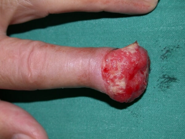
| Skin cancer destroying the finger tip |
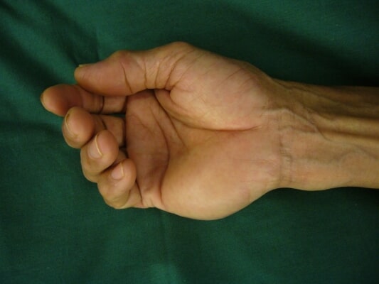
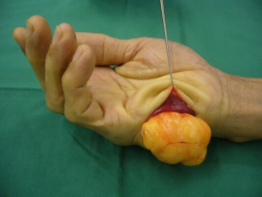
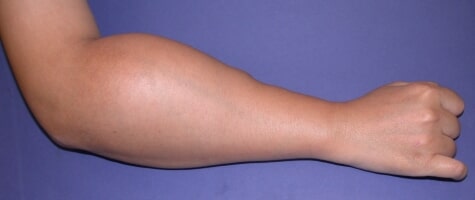
The commonest soft tissue tumours in the hand are fat tumours (lipoma), synovial tumours (pigmented villonodular synovitis), nerve tumours (neurilemmoma), and blood vessel tumours (vascular malformation). These are all primary tumours arising in the hand. Skin tumours are usually squamous cell carcinoma or melanoma, which are malignant.
|
|
|
| Fat tumour (lipoma) of the hand. | |
|
|
|
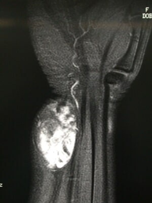
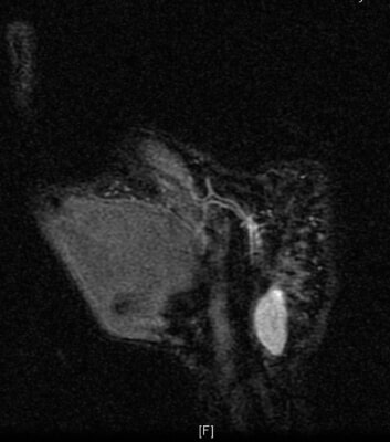
X-rays, ultrasound and MRI help to differentiate between the different types of tumours. Most of these tumours may be treated with excision biopsy. However, for large tumours, or tumours with a suspicion of malignancy, a biopsy should be done first before wide excision. Malignant tumours often require further tests, including bone scan, chest CT-scan, and PET scan to check the stage of the tumour. Where necessary, adjuvant treatment with chemotherapy may be necessary.
|
|
|
| A vascular malformation (blood vessel tumour) and a neurilemmoma (nerve sheath tumour) as shown on MRI. | |
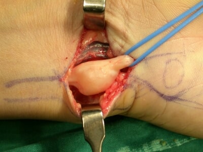
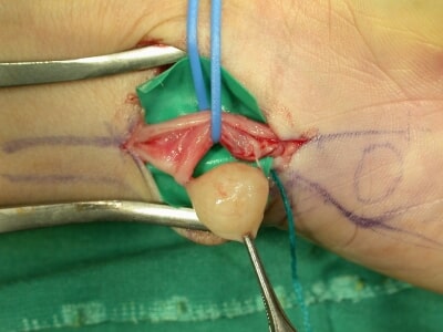
Nerve sheath tumours (neurilemmoma) of the nerves of the hand usually arise from a small branch of the nerve, but causes compression of the rest of the normal nerve. Treatment is excision of the tumour under the microscope, although this may occasionally result in some deficit of the nerve function.
|
|
|
| Neurilemmoma along the ulnar nerve, carefully dissected from normal nerve bundles. | |
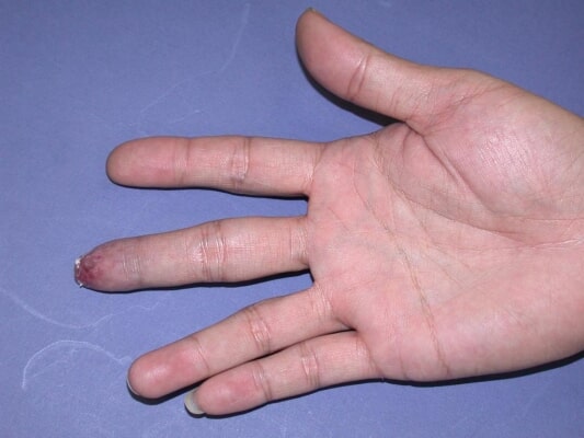
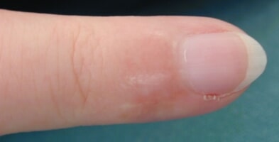
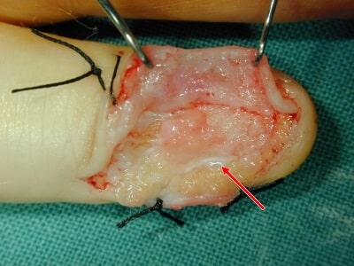
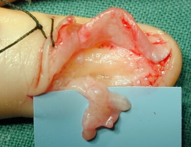
There is a peculiar tumour involving specialised cells of the blood vessels in the fingertip, usually at the nailbed, called a glomus tumour. It is very small, but causes a lot of pain, particularly when exposed to cold. It is difficult to diagnose, and a high index of suspicion is required. Ultrasound or MRI can help in the diagnosis. Complete excision is curative.
|
|
|
| Pain and swelling under nailbed, MRI showing vascular tumour in little finger | |
|
|
|
| Intra-operative findings of glomus tumour under nail bed. | |
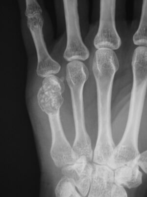
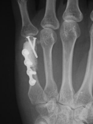
The commonest bony tumour is enchondroma, a cartilage tumour forming within the marrow cavity of the finger bone, resulting in weakening of the bone structure. The tumour itself is painless, but patients often present to the Accident & Emergency
Department or Hand Surgeon for acute pain after trivial trauma. The cause is fracture of the weakened bone due to a minor knock or twist. Treatment is to curette out the tumour, bone grafting and fixation of the fracture with plate and screws.
|
|
|
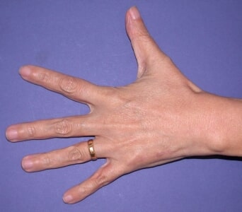
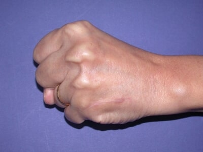
| Enchondroma usually present with pain due to pathological fracture, treated with excision, bone grafting and plate and screw fixation, | |
|
|
|
| With good outcome. | |
Secondary spread of a tumour arising from other sites are uncommon in the hand. The commonest cause is lung cancer. This often causes a fungating tumour of the finger. Local treatment is amputation of the finger. Management includes treatment of the primary source of the cancer.


